Neonatology
Session: Neonatal Infectious Diseases/Immunology 3
628 - Risk of Long-Term Neurological Sequelae in the “Asymptomatic” Congenital Cytomegalovirus Infection
Friday, May 3, 2024
5:15 PM - 7:15 PM ET
Poster Number: 628
Publication Number: 628.595
Publication Number: 628.595

Igor Nestrasil, MD, PhD (he/him/his)
Associate Professor
University of Minnesota Medical School
Minneapolis, Minnesota, United States
Presenting Author(s)
Background: The current state of knowledge is that a-cCMV individuals are not at disproportionate risk of major cognitive impairment when compared to healthy children. Since newborn cCMV screening is not universal and mild cognitive symptoms in infancy can be overlooked, a-cCMV infections may remain undetected in the early postnatal period or even later in life. On brain magnetic resonance imaging (MRI), cerebral white matter abnormalities showed in some a-cCMV infants. We employed a diffusion MRI (dMRI) technique sensitive to white matter integrity to quantify CMV impact on the white matter microstructure in a-cCMV infants.
Objective: Identify the impact of asymptomatic congenital cytomegalovirus (a-cCMV) infection on neurodevelopment assessed by brain dMRI in infants identified in the context of universal newborn screening.
Design/Methods: In this prospective longitudinal study approved by the University of Minnesota IRB, we enrolled and successfully acquired brain MRIs in 15 a-cCMV infants,13.13 ± 1.47 months old (8 females). MRIs were collected using the Baby Connectome Project (BCP) MRI protocol during physiologic sleep and compared to 26 typically developing infants whose data was derived from the BCP, 13.09 ± 1.34 months (15 females). Data were preprocessed, fitted with three diffusion models (diffusion tensor imaging, DTI; ball and 3 sticks, and neurite orientation dispersion and density imaging, NODDI), and reconstructed using highly reproducible deterministic tractography to yield averaged tract-specific microstructural and morphological measurements. Between-group differences were tested with the Wilcoxon rank-sum test.
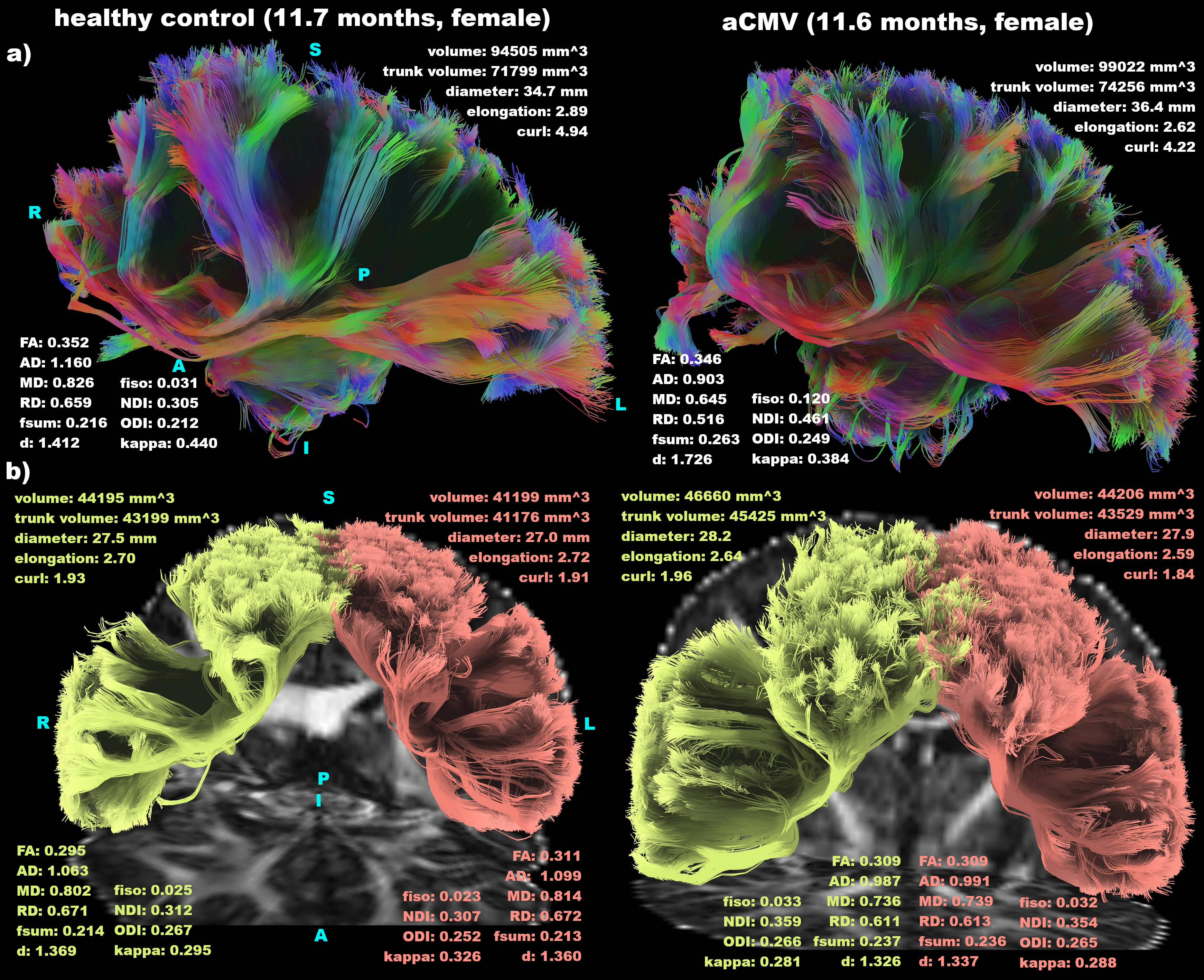
Results: Microstructural and morphological deficits were in corpus callosum forceps minor, frontal aslant tract (Table and Figure), corticostriatal tract, inferior fronto-occipital tract, superior longitudinal fasciculus 2, and anterior thalamic radiation.
Conclusion(s): We noted differences in dMRI between the a-cCMV and BCP infants. All affected tracts project to the frontal cortex, which suggests that the frontal lobe is the most profoundly affected structure in a-cCMV. Functionally, these tracts are involved in executive function, speech initiation, verbal fluency, goal-directed/motivated behaviors, impulse control, social behavior, emotional reactions, and attention in spatial orientation. All these functions are difficult to assess in infants, which may explain limited to no findings in the standard neurocognitive testing, resulting in “asymptomatic” cCMV manifestation. A larger dataset followed longitudinally beyond infancy is warranted to confirm these findings.


