Neonatology
Session: Neonatal Neurology 4: Clinical
532 - Development of Brain Functional Connectivity across Sleep States in Preterm Infants
Saturday, May 4, 2024
3:30 PM - 6:00 PM ET
Poster Number: 532
Publication Number: 532.1531
Publication Number: 532.1531

Anna Shiraki, MD (she/her/hers)
Visiting researcher
Nagoya University Graduate School of Medicine
Nagoya, Aichi, Japan
Presenting Author(s)
Background: The development of brain functional connectivity (FC) during active sleep (AS) and quiet sleep (QS) in preterm infants remains unclear. Furthermore, according to our preliminary study, FC analysis may be influenced by specific breathing patterns, such as periodic breathing (PB).
Objective: We assessed the impact of PB on FC analysis and explored the developmental trajectories of sleep state-dependent FC, excluding the influence of PB. Additionally, we investigated differences in FC associated with clinical characteristics using simultaneous electroencephalography and functional near-infrared spectroscopy data from preterm infants.
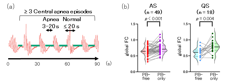
Design/Methods: We collected 148 records from 63 preterm infants (gestational age < 35 weeks) aged 31–40 weeks postmenstrual age (PMA), with a median recording duration of 68.5 min. Electroencephalography was performed polygraphically to distinguish AS from QS. PB was defined based on the American Academy of Sleep Medicine Manual guidelines (Fig. 1a). A portable 8-channel near-infrared spectroscopy device was positioned around the head to measure changes in the oxy-hemoglobin concentration. FC was calculated during AS and QS using slow fluctuations ( < 0.1 Hz) for segments lasting > 2 min. Global FC was determined as the average FC value across 28 pairs for PB-only and PB-free segments. Next, these pairs were categorized into five connection types (Fig. 2). The mean FC for each connection type during AS and QS, excluding PB segments, was assessed in relation to PMA. Additionally, FC data at 37–40 weeks PMA (n = 42) was used to investigate differences in mean FC according to gestational age, sex, and developmental quotient (DQ) at a corrected age of 10 months (low DQ [ < 85] or normal DQ [≥ 85]).
Results: Of the 148 recorded cases, 103 (70%) exhibited PB, regardless of PMA. Notably, PB-only segments exhibited significantly higher global FC in AS and QS compared with PB-free segments (Fig. 1b). The mean FC during AS increased with PMA, except for frontal-temporal connections, whereas the mean FC during QS did not change with PMA (Fig. 3a). At 37–40 weeks PMA, the mean FC in frontal-temporal connections during AS was higher in the low DQ than normal DQ group (Fig. 3b); no differences were observed according to gestational age or sex.
Conclusion(s): PB significantly distorts FC analysis in infants. Notably, distinct FC developmental patterns were observed between AS and QS at 31–40 weeks PMA. These findings are pivotal for understanding brain development in preterm infants.

.png)
.png)
