Neonatology
Session: Neonatal Neurology 6: Clinical
552 - White matter microstructure at 1 month is associated with the development of visuospatial processing.
Saturday, May 4, 2024
3:30 PM - 6:00 PM ET
Poster Number: 552
Publication Number: 552.1472
Publication Number: 552.1472
- NM
Narmin Mukhtarova, MD (she/her/hers)
Resident
University of Wisconsin Hospitals and Clinics
Madison, Wisconsin, United States
Presenting Author(s)
Background: Visuospatial processing is a non-verbal cognitive ability that operates upon perceptual stimuli and mental images and allow individuals to interact with their environment. Evidence shows that visuospatial processing and memory is present at 6 months and early recognition of the delay and initiation of supportive measures is effective
Objective: To determine if early white matter microstructure is associated with visuospatial abilities development
Design/Methods: Infants had MRI during non-sedated sleep at 1 month and behavioral assessment at 6 months. Diffusion tensor imaging (DTI) was used to calculate fractional anisotropy (FA) and radial diffusivity (RD) and neurite orientation dispersion and density imaging (NODDI) to calculate intracellular volume fraction (ICVF). Visual and sensorimotor white matter networks were chosen for analysis. “Puppet Task” was used to assess presence of startle, gaze shift for < 3 sec. and duration of visual engagement with two hand puppets. “Quilt” (baby activity pad) was used to assess changes in toy play and intensity of toy manipulation. Shapiro-Wilk, Student t-test, ANOVA and linear regression were used for statistical analysis
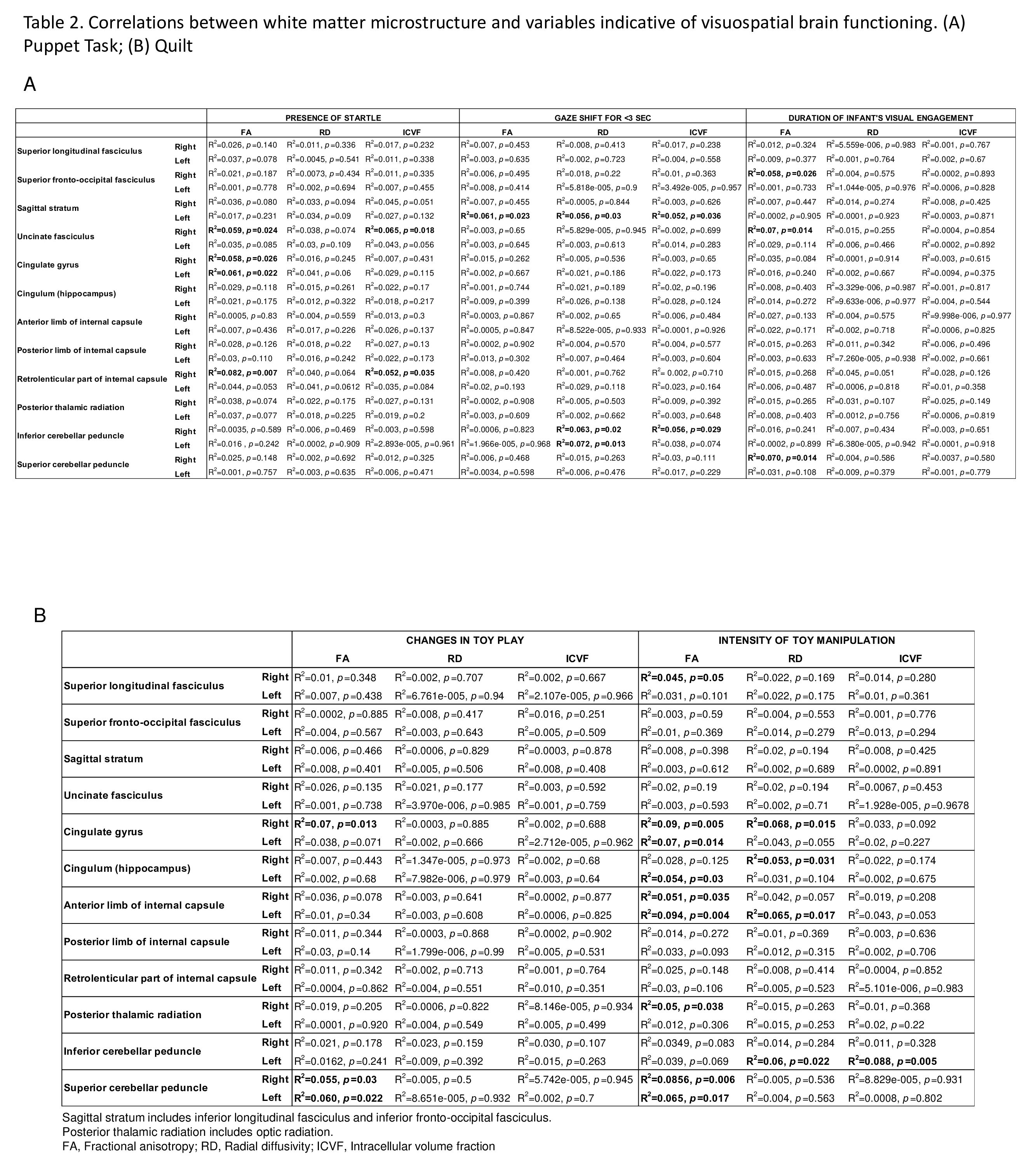
Results: Ninety-one infants were studied. Demographics are shown in Table 1. Negative correlations were found between: 1) presence of startle and FA in the right (R) uncinate fasciculus (UF), R & left (L) cingulate gyrus (CG), and R retrolenticular part of internal capsule (RLIC) and ICVF in the R UF and R RLIC (Table 2A); 2) gaze shift for < 3 sec and FA in the L sagittal stratum (SS) and ICVF in the L SS and R inferior cerebellar peduncle (ICP); 3) duration of engagement and FA in the R superior fronto-occipital fasciculus and R UF, and R superior cerebellar peduncle (SCP); 4) intensity of manipulation and RD in the R CG, R Cingulum (C), L anterior limb of internal capsule (ALIC), and L ICP (Table 2B). Positive correlations were noted between: 1) gaze shift for < 3 sec and RD in the L SS and R&L ICP; 2) changes in toy play and FA in the R CG and R&L SCP; 3) intensity of manipulation and FA in the R superior longitudinal fasciculus, R&L CG, L C, R&L ALIC, R posterior thalamic radiation, R&L SCP, and ICVF in the L ICP. Stratification of the intensity of manipulation data for male vs female infants showed higher rate of significant correlations in males compared to females (Table 3)
Conclusion(s): White matter microstructure of newborns may play a role in the development of visuospatial abilities. A more sustained correlations with widespread white matter in male infants are indicative of the effects of biological sex to the brain microstructure
Table 1. Demographics.jpeg

Table 3.jpeg
