Neonatology
Session: Neonatal General 6: POCUS, Technology in NICU
150 - Modified point of care ultrasound protocol for improving accuracy in confirmation of feeding tube position in neonates in a level III NICU
Sunday, May 5, 2024
3:30 PM - 6:00 PM ET
Poster Number: 150
Publication Number: 150.2146
Publication Number: 150.2146

Sfurti Nath, MD,FAAP
Assistant Professor of Pediatrics
University of Florida College of Medicine-Jacksonville
Jacksonville, Florida, United States
Presenting Author(s)
Background: Point of care ultrasound (POCUS) is essential to radiation conscious care. In addition to improving diagnostic and procedural accuracy, it can be effective in reducing cumulative radiation exposure. Researchers continue to explore newer and safer protocols for various POCUS applications. Our NICU created and adopted a POCUS protocol for confirmation of feeding tube (FT) position using a single epigastric tilted view. Due to limitations of static images, air interposition, movement artefact and non-visualization of the tip of FT, a number of false negative interpretations of FT position were observed. To improve accuracy of our protocol, we modified our approach by incorporating a dynamic video clip capturing a fluid flush to confirm presence of FT tip within the stomach and compared the two views.
Objective: To compare two imaging protocols for confirmation of FT position in neonates- static epigastric view and epigastric view along with dynamic video clip of fluid flush in sagittal epigastric view.
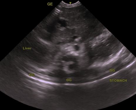
Design/Methods: Prospective observational study done in a level III NICU. Clinically stable infants ≥30 weeks GA on ≤ CPAP +6 were included. FTs were placed and confirmed per unit protocol. Clinician trained in POCUS and blinded to the results of x-ray obtained two images-one epigastric tilted view and one dynamic video clip capturing 2 ml fluid flush in epigastric sagittal view using GE venue 50 (curved array 4-8 MHz). Epigastric view identified FT as two echo bright parallel lines crossing into the stomach. Epigastric dynamic US noted air bubbles/ disturbance of liquid contents of the stomach on flushing 2 ml fluid through the FT indicating presence of FT tip within the stomach. The results of the two imaging protocols were compared to x-rays obtained ≤ 24 hours prior.
Results: n= 25, GA 33.6 (± 2.8) wks, mean BW 2068 (±840)grams. Time to obtain/interpret images was 45 (±9.8)secs for epigastric view and 1.8 (±2.3) mins for dynamic video clip with fluid flush. Mean drop in temp for both was 0.2C (±0.16).16% poor quality images obtained in epigastric view Vs 4% in dynamic video+ epigastric. 2/25 images in epigastric protocol failed to identify FT (false negative) giving a sensitivity of 91.6%. PPV was 100% and NPV was 33%. Whereas the dynamic video protocol had a sensitivity of a100% when compared to x-rays and a PPV and NPV of 100%.
Conclusion(s): Two view POCUS protocol incorporating epigastric view and dynamic video clip of fluid flush improved accuracy in confirming feeding tube position in neonates compared to single epigastric view. Larger studies are needed to assess generalizability of our results.

.jpg)
