Neonatology
Session: Neonatal Pulmonology - Clinical Science 5: New-Old ideas, Stem Cells
579 - Characteristics of Placental-derived Small Extracellular Vesicles (EVs) from Full term Healthy Pregnancies
Monday, May 6, 2024
9:30 AM - 11:30 AM ET
Poster Number: 579
Publication Number: 579.2744
Publication Number: 579.2744
.jpg)
Said Omar, MD
Professor and Division Chief of Neonatology
Michigan State University College of Human Medicine
Lansing, Michigan, United States
Presenting Author(s)
Background: Small Extracellular vesicles (sEV) are small membrane-enclosed vesicles that play a major role in immune modulation. Umbilical cord blood plasma (CB), Wharton Jelly (WJ) and placenta (Pl) mesenchymal stem cells (MSCs) supernatant are abundant source of EVs and may have a therapeutic effects in preventing acute lung injury. The characteristics of sEV derived from cord blood plasma (CB sEV), Wharton jelly (WJ sEV) and Placenta (Pl sEV) have not been extensively studied.
Objective: To examine the characteristics of CB, WJ and Pl derived sEV from healthy full term pregnancies.
Design/Methods: Placenta and Cord blood were collected after IRB approval and obtaining maternal informed consent. EVs were extracted from CB and supernatants of WJ and Pl MSCs culture. MSCs markers were analyzed by flow cytometry. The size, morphology and surface markers of sEVs were analyzed by Nanoparticle Tracking Analysis (NTA) and Transmission Electron Microscopy (TEM). and western blotting technique. Particle numbers, protein, and RNA amount of sEVs were measured. Biological effects of the extracted sEVs were tested on MDA-LUC cell proliferation, and on restoration of A549 and THP-1 cell viability when challenged with E-Coli derived lipopolysaccharide ( LPS).
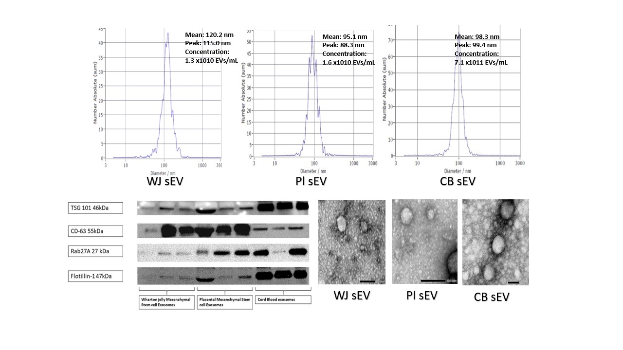
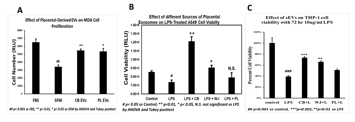
Results: Flow cytometry of isolated cells showed >90% expression of MSCs positive markers (CD73, CD90, CD44, CD105), < 1% of negative markers (CD34, CD31, CD11b), and stem cell markers (OCT4; 4-18%) and (SOX2; 70-80%).The isolation process of CB EVs was completed in 24 hours, while isolation of WJ and Pl sEV were completed in 3 weeks. CB sEV particles number was higher than Pl and WJ sEV (Table). The protein concentration was higher and the RNA content was lower in CB sEV compared with WJ and Pl sEV (Table). sEV characteristics is shown in Figure 1. CB, WJ and Pl sEVs promoted significant MDA cell proliferation (Figure 2A). CB and WJ sEV showed significant improvement in the viability of A549 lung epithelial cells and THP-1 cells by preventing LPS-induced cells injury (Figure 2B and 2C).
Conclusion(s): Cord blood Plasma, Placenta and Wharton Jelly derived small extracellular vesicles may have a promising therapeutic effect in the treatment of acute lung injury. The use of placenta as autologous source of small extracellular vesicles has the advantage of being rapidly isolated and may have large quantities to prevent lung injury in premature neonates.


.png)
