Neonatology
Session: Neonatal Neurology 6: Clinical
563 - Variations in cerebral hemodynamics in healthy infants measured continuously by trans-fontanel Doppler
Saturday, May 4, 2024
3:30 PM - 6:00 PM ET
Poster Number: 563
Publication Number: 563.1574
Publication Number: 563.1574

Astrid Ustad, MSc (she/her/hers)
Phd student
Norwegian University of Science and Technology
Trondheim, Sor-Trondelag, Norway
Presenting Author(s)
Background: Doppler ultrasound represents the gold standard for assessing cerebrovascular status in infants, providing a real-time overview of cerebral hemodynamics. However, conventional handheld Doppler studies offer only a “snapshot” status not reflecting the true temporal variation of cerebral blood flow.
Objective: The aim of this study was to describe physiological cerebral blood flow velocity (CBFV) variations in healthy infants in different states. In addition to a visual description, we aimed to present the continuous Doppler data numerically by statistical methods.
Design/Methods: NeoDoppler is a new monitoring device that provides real-time continuous cerebral Doppler assessment (Fig. 1). In this observational study, healthy term-born infants were recruited. A NeoDoppler probe was attached over the anterior fontanel (Fig. 1A). CBFV was measured in a cylindrical area below the probe (Fig. 1B-C). A validated quality indicator selected only high-quality Doppler spectra for analysis. Data was categorized based on state (sleeping, awake/active, breastfeeding) and postnatal age. Continuous data were analyzed using the means for each one-minute epoch when the infant remained in the same state. The effect of state on CBFVs was analyzed using linear mixed models. To describe temporal variations beyond those related to state, intraclass correlation coefficient (ICC) for two subsequent one-minute measurements (ICC short) and for two measurements 20 minutes apart (ICC long) were computed (adjusted for state).
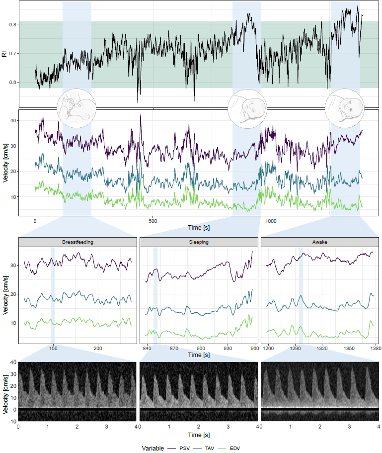
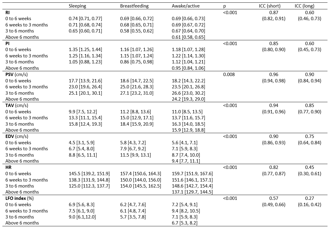
Results: Doppler data from 40 healthy infants (23 girls, postnatal age 1-48 weeks) were analyzed. The median recording time was 21.7 (range:1.6-30.0) minutes, resulting in valid measurements from a total of 99253 heartbeats (82.5%). A significant effect of state was observed in all Doppler variables (Tab. 1). Variations over time (ICC) beyond the effect of state were also seen inn all variables, being most prominent for the flow indices 20 minutes apart. CBFVs increase, while flow indices decrease with age, consistent with previous studies. Fig. 2 shows substantial variation in the Doppler parameters for one infant in different states when CBFVs were averaged over five heartbeats.
Conclusion(s): CBFV in infants is significantly influenced by their state and also changes substantially over time due to other factors. Prolonged Doppler acquisitions are necessary to accurately describe CBFV variations. This is important to be aware of using handheld ultrasound and when differentiating between normal variations and changes in patients at risk of cerebral blood flow compromise.


