Neonatology
Session: Neonatal Infectious Diseases/Immunology 3
620 - Clinical and pathological characteristics of a retrospective five year cohort of human cytomegalovirus infected placentas
Friday, May 3, 2024
5:15 PM - 7:15 PM ET
Poster Number: 620
Publication Number: 620.546
Publication Number: 620.546

Nitin Arora, MD MPH (he/him/his)
Assistant Professor
University of Alabama School of Medicine
Birmingham, Alabama, United States
Presenting Author(s)
Background: Congenital cytomegalovirus (CMV) is the most common congenital infection, affecting 0.5-2% of neonates delivered each year and leading to a large number of neonates requiring long-term medical care. The method of vertical transmission leading to significant effects continues to be studied.
Objective: Our objective is to characterize morphological changes in the placenta secondary to hCMV infection and evaluate the cell types infected and gene pathways involved using a novel spatial transcriptomic technique.
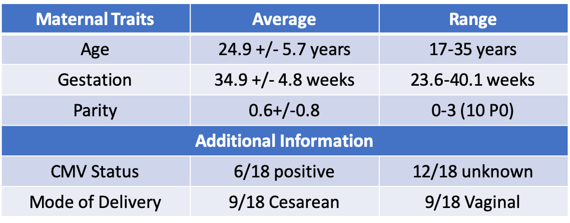
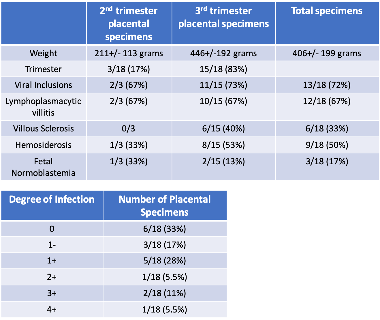
Design/Methods: From May 2018 to March 2023, there were 25 placental specimens obtained for evaluation, of which 18 were concerning for hCMV infection. The pathology of these placental samples was reviewed to identify characteristics that were commonly seen in neonates found to be hCMV positive. The placental pathology was then reviewed for all specimens to determine the presence of these features. Immunohistochemistry, immunofluorescence, and in situ hybridization techniques were then utilized to identify hCMV infection and replication products within placental specimens. Samples were then selected for transcriptomic evaluation based on cell counts and histopathologic findings to assess changes in biologic pathways amongst samples.
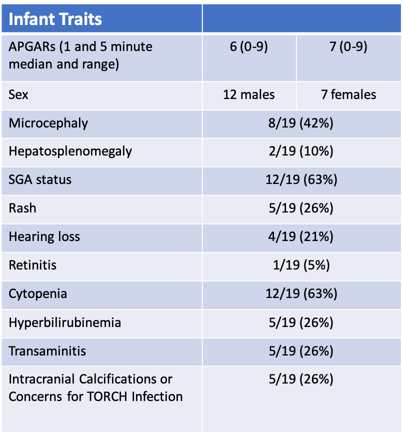
Results: In total, there were 19 infants that tested positive for CMV after delivery. 74%(14/19) of these infants had clinical findings based on examination and laboratory or imaging data. The placental samples were evaluated using techniques of immunohistochemistry, immunofluorescense, and in situ hybridization and the number of affected cells in each stained sample was assessed. A set of samples was identified to then perform spatial transcriptomic evaluation. The results of spatial transcriptomic evaluation are currently pending.
Conclusion(s): This project identifies a set of key morphological placental changes that are most notably associated with CMV infection and correlate with clinical disease. Novel techniques of immunohistochemistry, immunofluorescence, and in-situ hybridization, were then utilized to study infection within samples and to perform spatial transcriptomics for hCMV within infected placental specimen.


