Nephrology
Session: Nephrology 1
7 - Medical renal disease? pilot study on quantitative ultrasound of renal echogenicity
Saturday, May 4, 2024
3:30 PM - 6:00 PM ET
Poster Number: 7
Publication Number: 7.1087
Publication Number: 7.1087

Laura S. De Leon Benedetti, MD (she/her/hers)
Research Scholar
Childrens Hospital of Philadelphia
Philadelphia, Pennsylvania, United States
Presenting Author(s)
Background: Ultrasound is one of the diagnostic tools often utilized when assessing acute kidney injury (AKI). After discarding genitourinary obstruction or major anatomical anomalies, renal bladder ultrasounds are limited to subjective analysis of renal parenchymal echogenicity and/or corticomedullary differentiation preservation.
Objective: In this pilot study, we proposed a novel quantification method of renal texture analysis based on the spatial intensity patterns of the kidney tissue to detect texture variations among children with kidney disease when compared to young healthy controls.
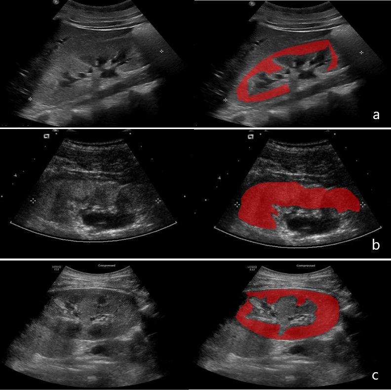
Design/Methods: We identified renal ultrasound images from 3 different groups: oliguric AKI (KDIGO stage 3 and not on dialysis), chronic kidney disease (CKD), and healthy controls. CKD cases and healthy controls were identified from NIH-approved prospective cohort studies (chronic kidney disease in children (CKiD) study, and Imaging Modalities in Pediatric Assessment of Kidney Transplants (IMPAKT) study, respectively). Texture analysis was conducted using the open-source software MaZda (version 4.6). The renal parenchyma was delineated, and various texture features were extracted, including first-order histogram, gray level co-occurrence matrix (GLCM), and run length matrix (RLM). These features were employed to evaluate the homogeneity and uniformity of the tissue's texture. Descriptive measures were calculated for each group. The median of each measurement was compared between the different groups using a Kruskall-Wallis test, and significant results were analyzed with the Conover test.
Results: A total of 31 subjects were analyzed: 8 AKI (F:6) with median age 3.5 years (IQR: 0-11.5), 14 CKD (F:6) with a median age of 3.5 years (IQR: 0-6.8), and 9 healthy controls (F:6) with median age 15.5 years (IQR: 12.8-21). Mean eGFRs (mL/min/1.73m2) at time of ultrasound for AKI and CKD subjects, and healthy controls were: 9.10, 61.98, 112.82, respectively.
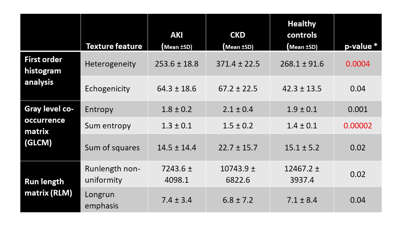
Several texture features were found to be statistically significant in distinguishing between the three groups (p < 0.05), Table 1. Mean sum entropy values from GLCM and mean heterogeneity from first-order histogram showed higher level of significance.
Conclusion(s): Conventional ultrasound texture analysis is a promising tool for understanding renal tissue changes in pediatric kidney disease, offering non-invasive insights into tissue composition. Significant differences in texture features between groups, as demonstrated in our study, underscore its clinical potential. Further research may lead to diagnostic and prognostic applications, improving patient care.


