Technology
Session: Technology 2
190 - Identification of Craniosynostosis from Non-Synostotic Skull Shapes with Volume Analysis
Sunday, May 5, 2024
3:30 PM - 6:00 PM ET
Poster Number: 190
Publication Number: 190.1942
Publication Number: 190.1942
- CK
Can Kocabalkanli, MSc
Computer Vision Research Engineer
PediaMetrix Inc
Rockville, Maryland, United States
Presenting Author(s)
Background: Craniosynostosis, characterized by the premature fusion of cranial sutures, impacts one in 2,000 births. If untreated early, it can lead to potential brain damage and often require surgical treatments. In contrast, deformational plagiocephaly or brachycephaly (DPB) which is non-synostotic, is prevalent in an estimated 20% of newborns and is managed with non-surgical measures like repositioning or helmet therapy. Early detection of both conditions presents a significant clinical challenge. The lack of efficient tools for pediatricians during regular visits leads to unwarranted referrals, treatment delays, and elevated medical costs.
Objective: To assist pediatric providers in the early identification and differentiation of synostotic and non-synostotic cranial conditions using head volumes, and facilitate early detection, objective referral, and intervention for both types of head deformity.
Design/Methods: Sets of 3D points were sampled from head volumes of 490 subjects (158 normal, 281 DPB, 51 craniosynostosis; mean age = 9 ± 11 months, median = 6 months; distributions illustrated in Fig. 1) as analysis entities. Data is collected by devices shown in Table 1. To address data imbalances, these point clouds were augmented with mirroring, resampling, and rescaling operations, with a total of 1088 shapes. Each shape was matched with and compared to a mean shape derived from 20 subjects with normal cranial conditions. The deviations underwent Principal Component Analysis (PCA) to identify key components as the bases of a constructed feature space. A Support Vector Machine (SVM) classifier was developed and evaluated using 3-fold cross-validation, during which the subjects were shuffled and split into “train” and “test” groups, and augmented data were built within its own group.
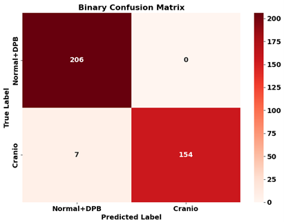
Results: The machine learning model classified cranial conditions as synostotic and non-synostotic with an average accuracy of 97.3 ± 1.1% accuracy, 94.6 ± 1.8% sensitivity, and 99.3 ± 0.7% specificity. Fig. 2 shows the confusion matrix of the binary classification on one-fold of unseen test dataset with 367 cranial shapes (52 Normal, 154 DPB, 161 craniosynostosis).
Conclusion(s): We devised a computational approach that differentiates 3D head volumes between craniosynostosis and DPB with notable precision. With statistical shape modeling and an SVM classifier at its core, this methodology presents an efficient screening tool, yielding 97.3% accuracy. Intended for pediatric settings, it has the potential to facilitate patient stratification when 3D scans are available, addressing the current gap in timely identification.


