Cardiology
Session: Cardiology 2
154 - Development of a Step-By-Step Freeware Solution for 3D Rendering of Congenital Heart Disease Images
Monday, May 6, 2024
9:30 AM - 11:30 AM ET
Poster Number: 154
Publication Number: 154.2934
Publication Number: 154.2934

Alan Groves, MD
Associate Professor
University of Texas at Austin Dell Medical School
Austin, Texas, United States
Presenting Author(s)
Background: 3D models of complex congenital heart defects show significant promise both for parent/provider education and for the process of surgical planning itself. However segmenting cardiac structures from CT or MR images is often labour intensive and can require expensive software platforms.
Objective: The goal of this study was to optimise and simplify the process of 3D rendering from CT images using freely available software.
Design/Methods: Example computed tomography (CT) and magnetic resonance (MR) imaging datasets were identified and downloaded in anonymised Digital Imaging and Communications in Medicine (DICOM) format. After reviewing a range of options, the free open-source software application 3D Slicer was deemed the most accessible and versatile for our needs. However, the National Institutes of Health (NIH) state that 3D Slicer is not intended for clinical use. A dedicated period of software feature discovery and application was combined with collaboration with the Center for Additive Manufacturing and Design Innovation (CAMDI) at the University of Texas at Austin to discuss different printing techniques.
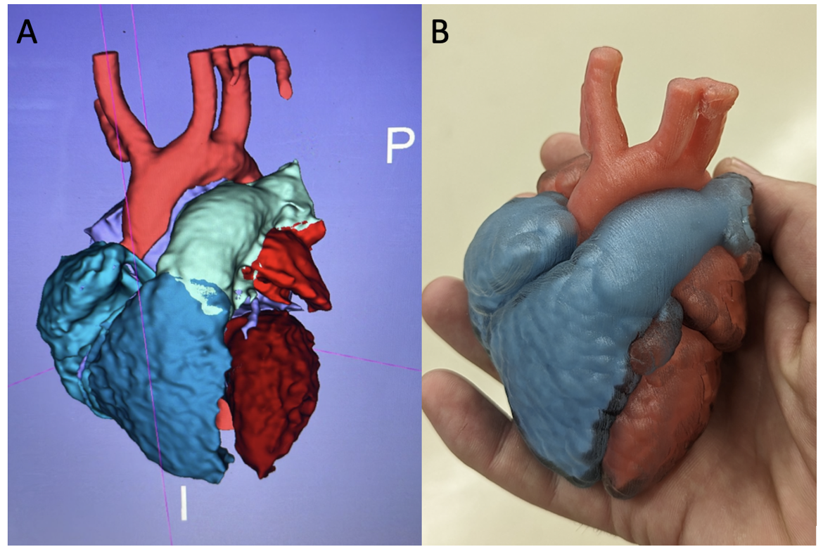
Results: The process of creating a replicable workflow for image segmentation culminated in the creation of a step by step guide which has been made freely available for download (https://utexas.box.com/shared/static/whqerjbl3kp3jylnxe029m4eyr0r8hvy.docx). Models consist of a colour-coded cardiac chamber blood pools (Figure 1A) which are stored as separate but linked .stl files. For 3D printing a solid blood pool can be encased in a softer myocardium shell with the model separating at the point of maximal anatomic interest for optimal viewing of internal defects (Figure 1B).
Conclusion(s): Following a simple, freely-available step-by-step guide and free software, CT and MR images of cardiac anatomy can be segmented into .stl file format. Such models can be rapidly and efficiently produced for visualization at the bedside, virtual reality exploration or for 3D printing.

