Newborn Care
Session: Newborn Care
223 - The prevalence of single umbilical artery in newborns and its associations with congenital anomalies
Friday, May 3, 2024
5:15 PM - 7:15 PM ET
Poster Number: 223
Publication Number: 223.533
Publication Number: 223.533

ibrahim Qattea, MD (he/him/his)
Fellow
Nassau University Medical Center
East Meadow, New York, United States
Presenting Author(s)
Background: A single umbilical artery (SUA) is a rare prenatal anomaly where a fetus has only one umbilical artery instead of the typical two. The condition is often detected during prenatal ultrasound or post-delivery examinations and has been associated with various neonatal anomalies. SUA occurs during fetal development, and while its exact cause remains uncertain, it is essential to understand its prevalence and potential associations with congenital anomalies and diseases.
Objective: This research aimed to evaluate the prevalence and trends of single umbilical artery (SUA) in infants born in the USA and investigate its association with chromosomal anomalies, congenital heart disease (CHD), congenital kidney disease (CKD), and congenital gastrointestinal disease (CGD).
Design/Methods: We utilized the de-identified national inpatient sample dataset from the Healthcare Cost and Utilization Project (HCUP) from the Agency for Healthcare Research and Quality (AHRQ). The study utilized a de-identified national dataset spanning from 2016 to 2020. The analysis incorporated all newborn inpatients with postnatal age ≤28 days. Patients were stratified based on sex, racial background and hospital bed size. The Cochran-Armitage trend test was implemented for trend analyses and regression analyses were performed to determine adjusted Odds Ratios (aOR).
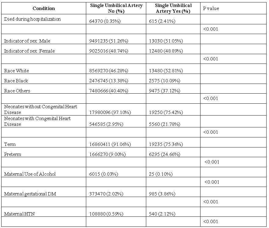
Results: Dataset included 18,916,425 newborns, 365,739 of them were excluded due to transfers to other centers, resulting in 18,550,686 infants included in the analysis. The total number of infants with SUA was 24910 (0.13%) of them 4,755 (0.9%) cases with CHD, 2,280 (1.64%) cases with CKD and 1,165 (1.83%) cases with CGD.
CHD showed a significant association (p-value < 0.001) with an adjusted odds ratio (aOR) of 8.13 (95% CI: 7.57 - 8.74). CKD and CGD were associated (p-value < 0.001) with aORs of 13.66 (95% CI: 12.18 - 15.32) and 14 (95% CI: 12.57 - 15.61), respectively. Down Syndrome was associated with SUA (p-value < 0.001) with an aOR of 4.18 (95% CI: 3.56 - 4.92). Trisomy 18 and Trisomy 13 also exhibited significant associations (p-value < 0.001) with aORs of 42.8 (95% CI: 36.36 - 50.36) and 28.33 (95% CI: 22.73 - 35.28), respectively. Finally, Trisomes displayed a significant association (p-value < 0.001) with an aOR of 8.97 (95% CI: 7.97 - 10.09).
Conclusion(s): SUA is associated with multiple chromosomal and congenital anomalies. The study may provide guidance for clinical practice when diagnosing an infant with SUA.


