Neonatology
Session: Neonatal Neurology 5: Clinical
545 - Functional near-infrared spectroscopy reveals altered resting-state connectivity in preterm newborns with intraventricular hemorrhage
Saturday, May 4, 2024
3:30 PM - 6:00 PM ET
Poster Number: 545
Publication Number: 545.1654
Publication Number: 545.1654

Lingkai Tang
PhD candidate
Western University
London, Ontario, Canada
Presenting Author(s)
Background: Intraventricular hemorrhage (IVH) is a major complication following preterm birth. IVH can lead to mortality or long-term motor and cognitive deficiencies. fNIRS can be a new bedside monitoring tool to image brain function in neonates with brain injury, through resting-state functional connectivity (RSFC).
Objective: To examine if fNIRS could differentiate between preterm born neonates with IVH and healthy controls, we compared RSFC patterns of the two groups using graph theory based measurements.
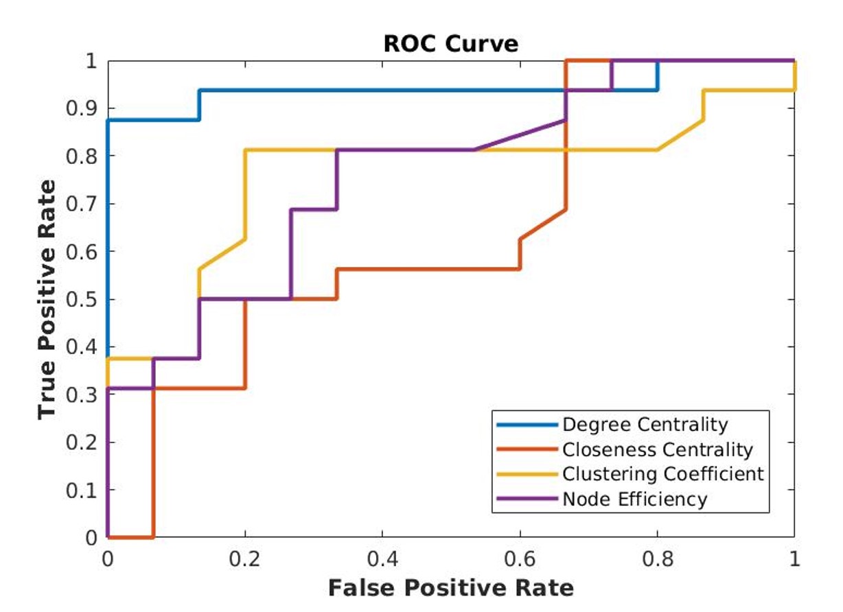
Design/Methods: fNIRS system (NIRSport2) was set up with 20 channels, covering the whole head. All neonates had a 6-min resting-state scan. IVH group included 16 preterm born neonates (gestational age [GA] at birth =26.3 weeks, postmenstrual age [PMA] at scan = 37.0 weeks) who were categorized into mild or severe group based on severity of their injuries. 15 healthy term-born neonates (GA at birth = 38.9 weeks) were also recruited. Cerebral hemodynamics was derived from fNIRS data from each channel then correlated with each other to create a RSFC map for each neonate. We measured the similarity of RSFC maps of mild or severe group, against no injury, respectively, by calculating the Euclidean and Jaccard distances. We then assessed the association between severity of IVH and connectivity strength using generalized linear models adjusted for GA, PMA and sex. Graph theory based measurements (clustering coefficient, network efficiency, etc.) were also used for calculating global connectivity patterns and local patterns. SVM classifiers were trained with connectivity features to distinguish IVH and control groups. Receiver operating characteristic (ROC) curves were plotted and areas under the curves (AUC) were calculated.
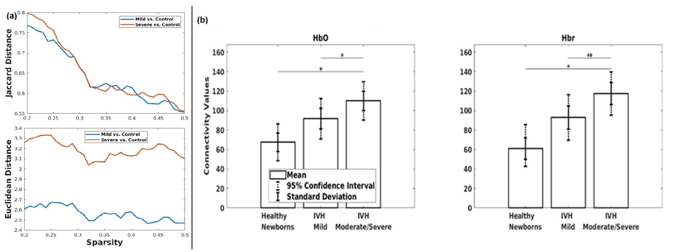
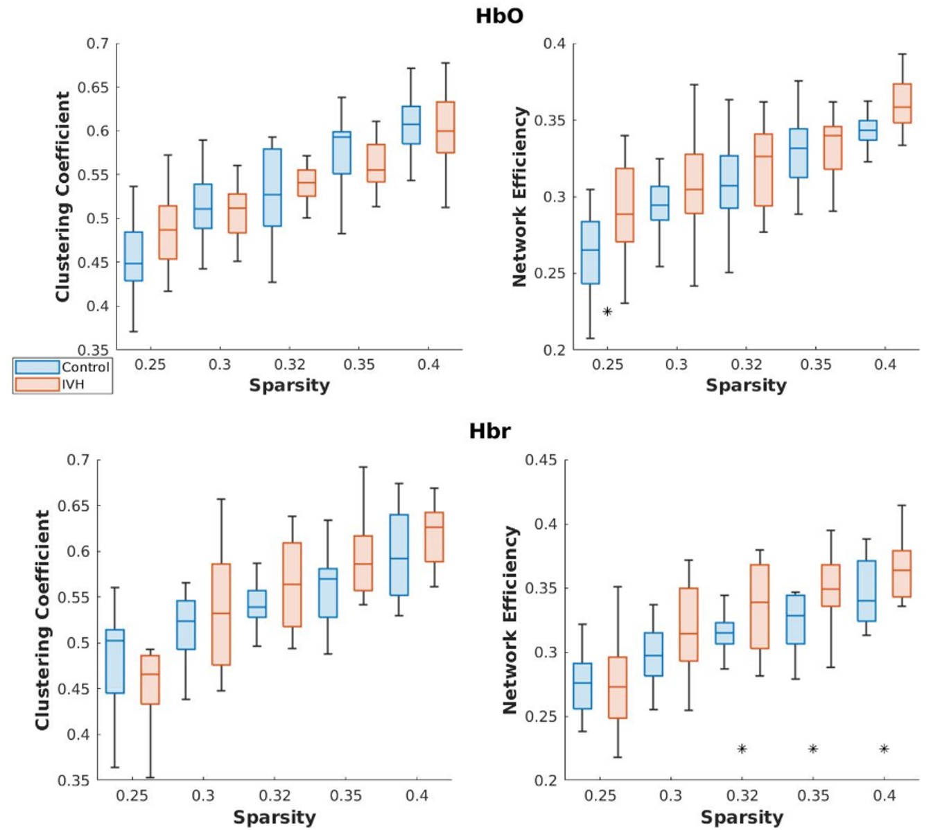
Results: Results showed that mild IVH group overlapped more with healthy control than severe group at different levels of sparsity (Fig. 1a). This indicates that functional organization of brain is less disrupted within mild-injury group. Connectivity strength increased with severity of injury (Fig 1b). IVH group showed increased clustering coefficient and network efficiency as well (Fig. 2). With the connectivity features, a maximal accuracy of 93.55% and AUC of 0.94 were achieved (Fig. 3).
Conclusion(s): In conclusion, fNIRS revealed differential RSFC patterns between groups of neonates with IVH and healthy controls. This indicates that RSFC derived from fNIRS can be a potential marker for severity of IVH. We showed that fNIRS can potentially be a new tool for assessing early brain injury and monitoring cerebral hemodynamics of newborns.


