Neonatology
Session: Neonatal Neurology 8: Preclinical
327 - Dose-Dependent Effect of Caffeine on Cell Death and Oxidative Stress in a Ferret Organotypic Brain Slice Model of Hypoxia-Ischemia is Partially Dependent on A2A Receptors
Monday, May 6, 2024
9:30 AM - 11:30 AM ET
Poster Number: 327
Publication Number: 327.3135
Publication Number: 327.3135

Olivia C. Brandon, BS (she/her/hers)
Research Scientist
University of Washington School of Medicine
Seattle, Washington, United States
Presenting Author(s)
Background: Brain injury after hypoxia-ischemia (HI) is the leading cause of neonatal morbidity and mortality worldwide. The ferret provides an excellent model in which to study HI due to its gyrified brain and white-to-gray matter ratio that more closely resembles humans compared to rodents. Caffeine is neuroprotective in rodent models of HI and in infants treated for apnea of prematurity, but the mechanisms of neuroprotection, including antioxidant and anti-inflammatory signaling via A2 adenosine receptor (A2AR) antagonism, have not been fully explored.
Objective: To assess caffeine’s effect on neuronal cell death, cytotoxicity, and inflammatory and oxidative stress markers in a ferret organotypic brain slice model of HI.
Design/Methods: We used an established ex vivo model of term-equivalent whole-hemisphere ferret brain slices that retain the cerebral architecture of in vivo models. After 72-hours in culture, 300 µm slices were exposed to oxygen-glucose deprivation (OGD) for two hours to mimic HI. Slices were randomized to OGD, OGD with caffeine (20mg/kg or 50mg/kg) and A2AR agonist (2mg/kg). Non-exposed healthy slices were used as controls. Analyses included lactate dehydrogenase (LDH) release assays, cell death indicated by pyknotic nuclei, reduced glutathione (GSH) and oxidized glutathione (GSSG) levels. qRT-PCR assessed gene expression related to oxidative stress (HMOX) and inflammation (IL10 and TNFa).
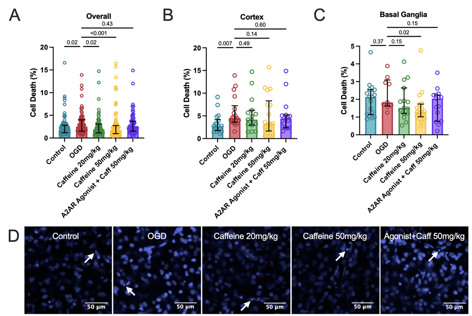
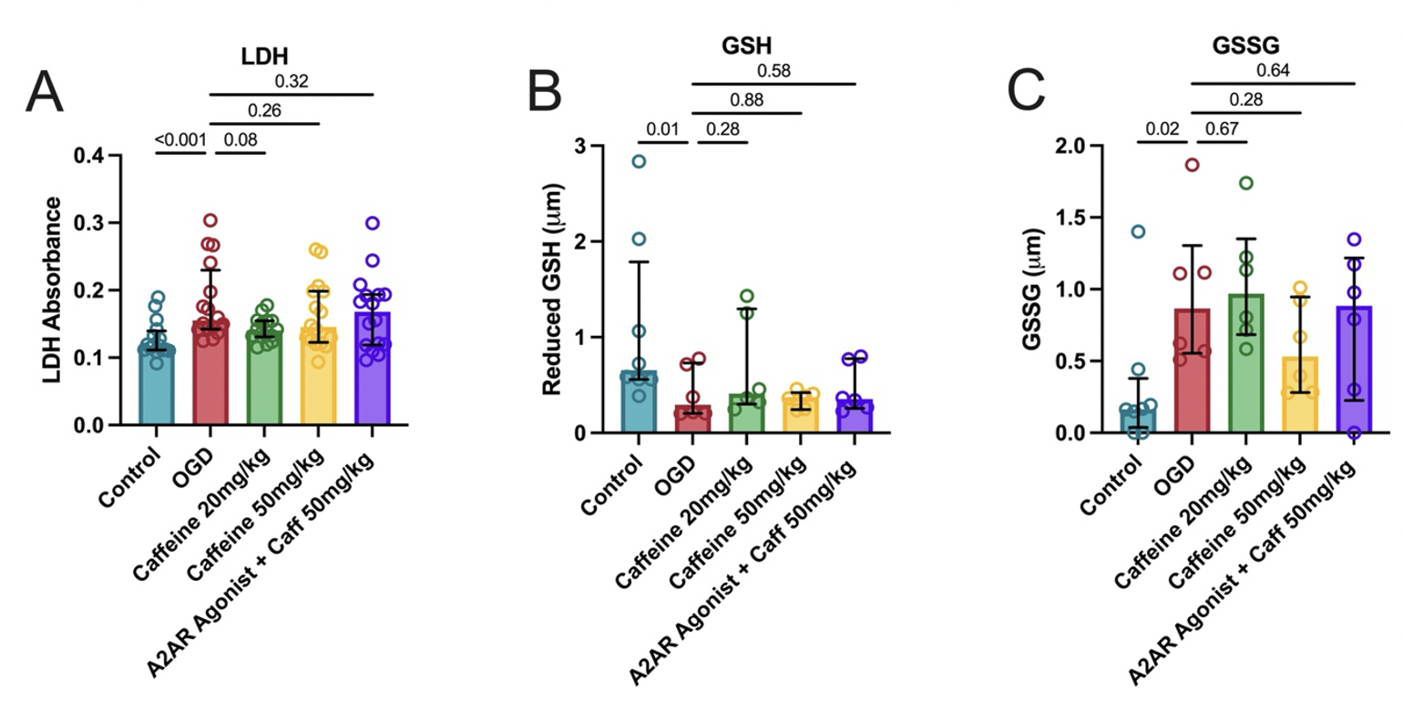
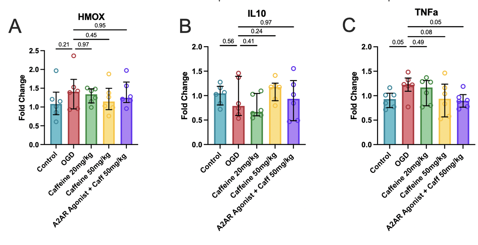
Results: Exposure to caffeine at 20 and 50mg/kg decreased cell death compared to OGD (p=0.02, p< 0.001; Fig 1A). Adding the A2AR agonist reversed caffeine neuroprotection (p=0.43 vs. OGD; Fig 1A). Similar regional trends were seen in the cortex (Fig 1B) and basal ganglia (Fig 1C), and cortical representative images (Fig 1D). OGD significantly increased global cell death, as measured by LDH absorbance, compared to controls (p < 0.001; Fig 2A) which was reversed by caffeine at 20mg/kg but not at 50mg/kg. OGD had lower GSH (p=0.01; Fig 2B) and higher GSSG (p=0.02; Fig 2C) compared to controls. Markers of oxidative stress and inflammatory were altered by OGD – increased expression of HMOX, TNFa and decreased expression of IL-1B – and were partially, but not significantly normalized by caffeine at 50mg/kg (Fig 3).
Conclusion(s): In this ex vivo ferret model of HI, caffeine decreased cellular death and decreased oxidative stress which was partially reversed with the A2AR agonist. Higher powered studies across a range of preclinical models are warranted to further explore the effects of caffeine on HI.


