Neonatology
Session: Neonatal Neurology 7: Neurodevelopment
613 - Brain Metabolite Concentrations in Neonates of Diabetic Mothers
Saturday, May 4, 2024
3:30 PM - 6:00 PM ET
Poster Number: 613
Publication Number: 613.1171
Publication Number: 613.1171

Nickie Andescavage, MD
Associate Professor
Children's National Health System
Washington, District of Columbia, United States
Presenting Author(s)
Background: Infants of diabetic mothers (IDM) suffer from a higher risk of metabolic disorders including hypoglycemia and hypocalcemia. Their etiologies and managements have been relatively well studied and documented. Investigations and treatments have been focused on conditions that affect overall metabolism but the brain biochemistry has been neglected in which only one proof-of-concept study has reported brain metabolite concentrations in fetus of diabetic mothers; there are no reports of similar studies on neonates.
Objective: The objective of this study was to compare in vivo brain biochemistry in neonates of diabetic mothers with those of non-diabetic mothers.
Design/Methods: Neonates of diabetic mothers were recruited prospectively. Informed written consents were obtained. Inclusion criteria for neonates included mothers with gestational or pregestational diabetes. Exclusion criteria included any contraindications of MRI, documented chromosomal abnormality, central nervous system anomalies and multiple gestations.
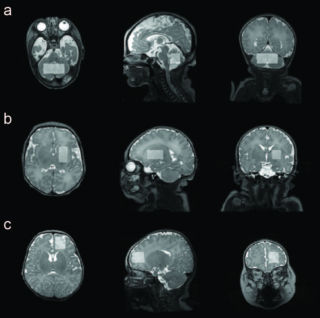
Clinical data for the neonates including gestational age (GA) at birth, weight at birth, sex, GA at MRI. All MRI scans were performed on a 3T scanner using an 8-channel surface receive coil during natural sleep without sedation. T2-weighted 3D CUBE FSE sequence was used to acquire anatomical images for voxel positioning. MR spectra were acquired using the MEGA-PRESS. Voxels were positioned in the cerebellum, right frontal lobe and right basal ganglia (Fig 1). Spectra were processed and modeled using the Osprey software v2.4. Metabolites included are reflected in Table 2. All statistical analyses were performed using R v4.3.1 in RStudio. Independent samples t-test was performed to compare metabolite measurements between groups.
Results: MRI and MRS were successfully obtained from 41 controls and 36 cases Table 1. Data with low SNR, excessive motion artifacts and lipid contamination were excluded. Independent sample t-test indicated that between cases and controls, metabolite measurements of tNAA, tCho and tCr were significantly different (P < 0.05) in the cerebellum; GABA was significantly different in right frontal lobe, as indicated in Table 2.
Conclusion(s): In this pilot study, we found significant differences in in vivo brain metabolites in IDM compared to healthy controls. For IDM, GABA in the right frontal lobe is significantly higher although animal studies suggest downregulation of GABA receptors in diabetes, which suggests this increase may be compensatory in nature. Additional differences in tNAA, tCho and tCr were found in the cerebellum, though needs to be validated in larger studies.

.jpg)
.jpg)
