Neonatology
Session: Neonatal Cardiology and Pulmonary Hypertension 4: Congenital Heart Disease
172 - In Utero Endothelial Cells Have Abnormal Shape and Under-Express Vasculature Development Genes in Single Ventricle Congenital Heart Disease
Monday, May 6, 2024
9:30 AM - 11:30 AM ET
Poster Number: 172
Publication Number: 172.2946
Publication Number: 172.2946

Angela Desmond, MD (she/her/hers)
Neonatal-Perinatal Medicine Fellow
UCLA Mattel Childrens Hospital
Santa Monica, California, United States
Presenting Author(s)
Background: Congenital heart disease (CHD) is the most common major birth defect and represents the leading cause of infant death due to a congenital anomaly, yet etiologies and developmental mechanism are not well defined. The umbilical cord offers a unique platform to study in-utero vasculature.
Objective: In this study, we hypothesized human umbilical vein endothelial cell shape and differentially expressed genes (DEGs) would be aberrant in pregnancies affected by single ventricle (SV) CHD physiology compared to controls.
Design/Methods: This was a prospective cohort study (IRB #19-001754) of pregnancies affected by fetal SV CHD (n=8 for cell shape, n=19 for RNAseq) and controls (n=6 for cell shape, n=84 for RNAseq). Umbilical cords were collected at delivery, processed, and exposed to collagenase to isolate venous endothelial cells. Cells were cultured and passaged. To mimic flow, cells were subjected to shear stress through an orbital shaker (130 rpm) or unidirectional flow by ibidi pump system. After staining, endothelial cell elongation factor (EF=Length/Width), shape index (SI=(4π*Area)/(perimeter^2)), and angle relative to flow were calculated. Two-tailed Mann-Whitney statistical testing was used. RNA extraction, library preparation, and sequencing were performed through RNeasy Micro Kit, TruSeq Total RNASeq Kit, and RNAseq. Benjamini-Hochberg false discovery rate (FDR) method was used with significance threshold of FDR < 0.05. DESeq2 was used to interpret differential gene expression results. GO biological processes performed gene ontology term enrichment of identified DEGs.
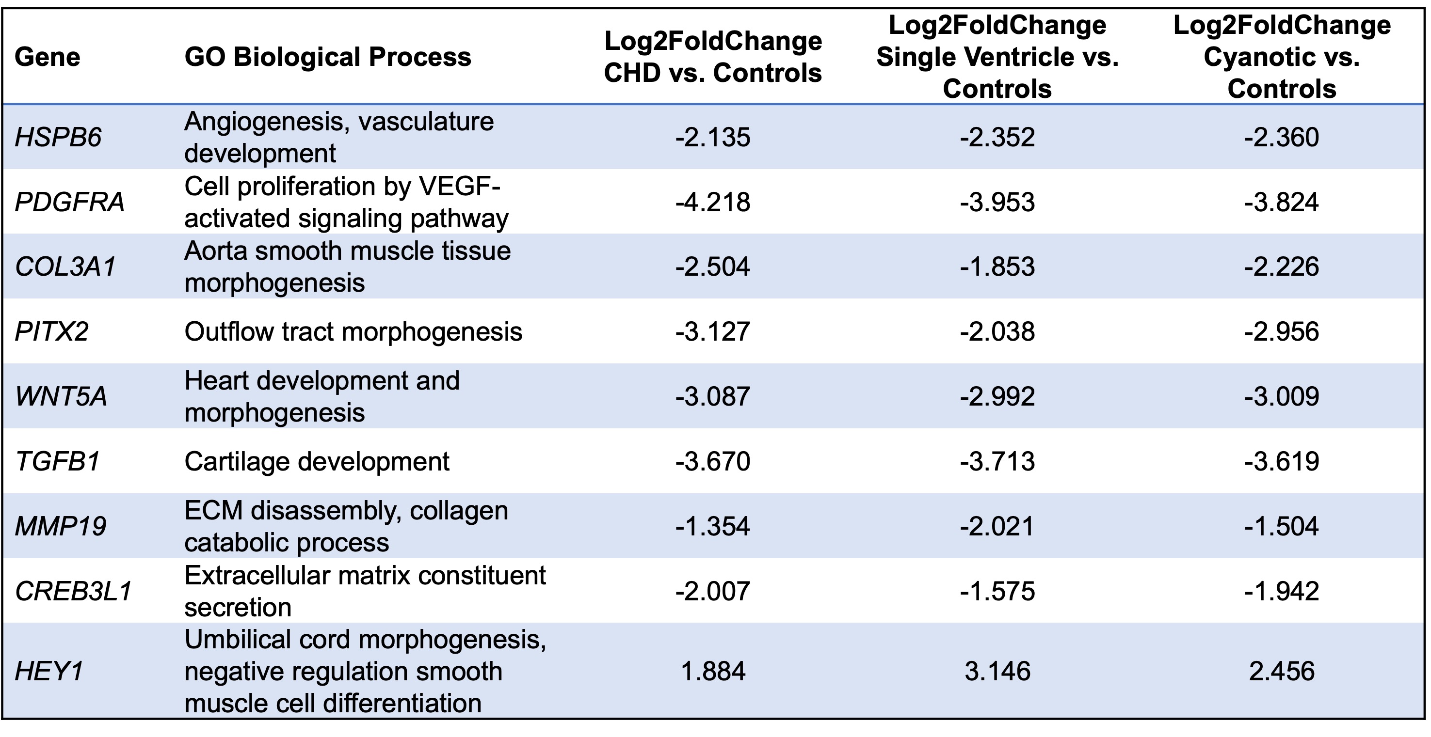
Results: Under flow, the SV cohort’s endothelial cells’ EF and angle relative to flow were significantly different from controls (p=0.02, p=0.05). There was no significant difference in SI (p=0.36). SV CHD cases compared to controls had 75 DEGs with notable under-expression of genes involved in vasculature development (HSPB6, PDGFRA, COL3A1), heart development (WNT5A, PITX2) and extracellular matrix organization (TGFB1, MMP19, CREB3L1).
Conclusion(s): When exposed to flow, umbilical cord endothelial cells from SV CHD pregnancies showed altered elongation compared to controls. Endothelial cells isolated from SV CHD pregnancies under-express genes involved in vasculature development and extracellular matrix organization. Abnormal vascular flow during development may contribute to these shape and gene expression findings. Additional studies are needed to investigate the broader hypothesis of an abnormal vascular phenotype predisposition for CHD.

